
SVT ECG findings: recognizing supraventricular tachycardia in children
SVT ECG findings can be easy to overlook in young children. This case-based lesson shows how key ECG features support the diagnosis of supraventricular tachycardia in a neonatal patient.

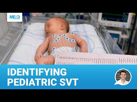
SVT ECG findings are the focus of this neonatal ECG case, where a very fast heart rate and hard-to-identify P waves make rhythm assessment tricky. In this case-based lesson, you’ll assess the ECG from a 3-day-old baby with narrow complex tachycardia and see how key ECG features support the diagnosis. Work through the rhythm step by step, then learn how an accessory pathway can create re-entrant SVT in infancy.
In this lesson from our Pediatric ECG course, you’ll learn how to:
- Assess rhythm on a neonatal ECG
- Identify hard-to-see P waves in pediatric SVT
- Use heart rate to evaluate tachycardia in a newborn
- Recognize narrow QRS complexes on ECG
- Interpret P waves that appear after the QRS complex
- Connect regular, narrow complex tachycardia with SVT
- Understand how an accessory pathway can create re-entrant SVT in infancy

Start the first chapter of our Pediatric ECG course for free
Supraventricular tachycardia in children
[0:00]
Supraventricular tachycardia, or SVT, is one of the most common arrhythmias in young children. It can turn a little heart into a racing engine without warning. This Medmastery lesson will show you how to detect the subtle ECG signs of SVT in pediatric patients, giving you the tools to spot this arrhythmia when it matters most.
In this lesson, we will visit the Neonatal Intensive Care Unit. Our patient is a 3-day-old baby in tachycardia. An ECG was obtained, but the rhythm assessment is a bit tricky for this patient.
P waves and SVT ECG findings in a newborn
[0:35]
At first glance, it is difficult to find any P waves, but a zoomed-in version shows a little tiny deflection after the QRS complex that might be a P wave. It is very difficult to see the P waves in this ECG, let alone determine if they're upright or inverted.
Assessing heart rate: supraventricular tachycardia in neonates
[0:52]
Let's move on to the rate. This ECG indicates a super fast heart rate of about 250 beats per minute. This is definitely not normal, not even for newborns, whose heart rates typically range from 100 to 160 beats per minute.
QRS axis and intervals in pediatric ECG interpretation
[1:07]
Next, the QRS axis is normal, with an upright QRS in leads I and aVF. Now let's look at the intervals. The PR interval is a bit tricky, since the P waves seem to be coming right after the QRS, making it difficult to evaluate them.
Narrow complex tachycardia in very young children
[1:24]
If we look at the QRS complexes, they appear narrow. Collectively, these findings, a narrow QRS and P waves appearing after the QRS complexes, indicate a regular, narrow complex tachycardia, a very common arrhythmia in the very young.
SVT diagnosis in a newborn
[1:41]
This baby has supraventricular tachycardia, or SVT. Supraventricular tachycardia, or SVT, is an umbrella term for many different arrhythmia mechanisms, all arising from the atria. It turns out that a regular, pretty fast, narrow complex tachycardia in newborns is almost always something called re-entrant supraventricular tachycardia.
Accessory pathways and the re-entrant SVT mechanism
[2:05]
Essentially the electrical conduction in the atria short circuits. Instead of simply traveling from the atria to the ventricles, an accessory pathway allows the electrical impulse to re-enter the atria, creating a loop or circuit. When this happens, the heart beats very fast. If left untreated and unrecognized, patients can develop more serious symptoms related to heart failure.
Accessory pathways in fetal heart development
[2:28]
Parents often wonder why their seemingly healthy baby developed a heart problem. The explanation lies in how the heart forms in utero. During early development, the atria and ventricles start out as a single unit. As the fetus develops, the atrium and ventricles slowly separate from one another, and during development, primitive conduction pathways initially connect the atria and ventricles. But most of these pathways disappear as the heart matures, leaving only the AV node and His-Purkinje system.
AV node development and the origin of pediatric arrhythmias
[3:00]
But sometimes a remnant conduction pathway persists as an accessory pathway, allowing a re-entrance circuit to occur even after birth. I use a pizza analogy to explain this. Imagine taking a piece of pizza. As you pull the pizza slice away, tiny strings of cheese remain attached. These strings of cheese are like the accessory pathways. In many babies, these extra connections will eventually go away on their own, but before they do, they can cause arrhythmias like they did in our patient.
Pediatric SVT treatment through infancy
[3:31]
Most babies with SVT in infancy will be treated through the first nine months of life or so, hoping that these connections just vanish on their own with continued heart development. That's exactly what we did for our patient, and after one year of life, she no longer had SVT.